
For Informational Purposes Only
Intraoperative Airway Management with a Tracheostomy Tube
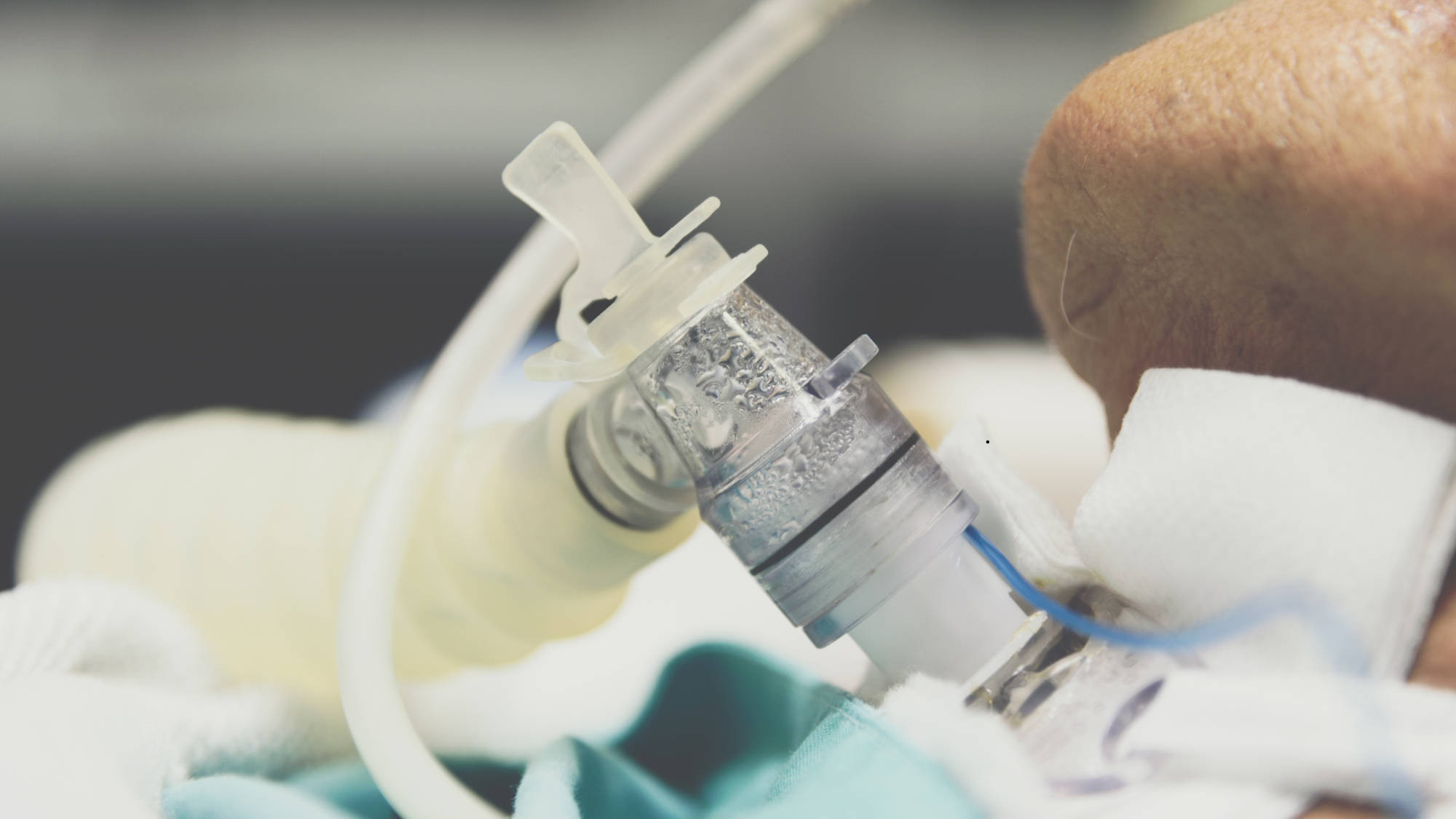
Patients who present with an upper airway obstruction, require mechanical ventilation, or need removal of trachea-bronchial secretions may require a tracheostomy. A tracheostomy involves surgically opening the area of the throat inferior to the larynx and inserting a curved rubber respiratory tube. A variety of different tracheostomy tubes are available, including ones with a 15mm extension, obturator, inner cannulas, or a cuff. Depending on patient history and condition, different add-ons may be more desirable.[i]
While intraoperative airway management with a tracheostomy tube can help mitigate respiratory crises, there are a number of considerations that should be taken into account during intubation. For example, children under the age of two have the highest rate of composite morbidity within 30 days following surgery at approximately 23 percent,[ii] and for this reason other airway management options should be considered. If the patient does not have any other viable options, then anticipation of complications and increased monitoring may be necessary.
One of the most dangerous complications of receiving an intraoperative tracheostomy is obstruction of the intubated airway which can lead to cardiorespiratory distress. Obstruction can occur for a variety of reasons, including the presence of thick secretions, twisting/kinking of the rubber tube, mucous plugs, blood clots, and foreign bodies. Accidental dislodgement of the tube is also a common reason for cardiorespiratory distress. There are several early warning signs of obstruction, including tachypnea, labored breathing, tachycardia, and decreased levels of oxygen saturation. Prolonged obstruction increases the risk of cyanosis, bradycardia, and apnea, all of which can result in long-term adverse effects.[iii] It is therefore highly preferable that physicians are able to identify and intervene in the early stages of obstruction as a result of consistent patient monitoring and close supervision of oxygen saturation levels.
Other complications that can arise from tracheostomies are unique to the first post-tracheostomy week. These include stomal erosion, respiratory and/or cardiovascular collapse, cellulitis, granulation tissue at the site of the insert, and tracheo-esophageal fistulas. Both in the first week and in the following weeks, there remains risk of infection.[iv] Care teams generally aim to keep the area of the incision as clean as possible to help manage this risk. Published guidance commonly describes once-daily dressing changes and stoma cleaning as a general baseline for wound care, though this can vary by patient and institutional protocol. Care should also be taken not to disturb the site of the insert: a study by Espinel et al. showed that patients with more than four manipulations per day in the two weeks following a tracheostomy were significantly more likely to have a complication.[v] Continuous pulse oximetry is commonly used for patients who are unconscious or not under direct observation by a caregiver, to help support awareness of oxygen levels. Having tracheal dilators available near the bedside, along with an information sheet noting details such as tracheostomy tube type/size and the date of the last tube change, are practices some care teams use to help support faster, more informed intervention in the event of a crisis.[vi]
In sum, intraoperative tracheostomies are often necessary for patients with compromised upper airways. Though the rate of tracheostomy-related complications is typically low, there are a number of considerations that can mitigate these risks even further, particularly for more vulnerable younger populations.
References
[i] Shah, R. K., Stey, A. M., Jatana, K. R., Rangel, S. J., & Boss, E. F. (2014). Identification of opportunities for quality improvement and outcome measurement in pediatric otolaryngology. JAMA Otolaryngology, 140(11), 1019–1026. https://doi.org/10.1001/jamaoto.2014.206
[ii] Mahida, J. B., Asti, L., Boss, E. F., Shah, R. K., Deans, K. J., Minneci, P. C., & Jatana, K. R. (2016). Tracheostomy Placement in Children Younger Than 2 Years: 30-Day Outcomes Using the National Surgical Quality Improvement Program Pediatric. JAMA Otolaryngology, 142(3), 241–246. https://doi.org/10.1001/jamaoto.2015.3302
[iii] Epstein S. K. (2005). Late complications of tracheostomy. Respiratory Care, 50(4), 542–549.
[iv] Mathisen D. J. (1996). Complications of tracheal surgery. Chest Surgery Clinics of North America, 6(4), 853–864.
[v] Espinel, A. G., Scriven, K., & Shah, R. K. (2019). Tracheostomy manipulations: Impact on tracheostomy safety. Pediatric Investigation, 3(3), 141–145. https://doi.org/10.1002/ped4.12141
[vi] Roxbury, C. R., Jatana, K. R., Shah, R. K., & Boss, E. F. (2017). Safety and postoperative adverse events in pediatric airway reconstruction: Analysis of ACS-NSQIP-P 30-day outcomes. The Laryngoscope, 127(2), 504–508. https://doi.org/10.1002/lary.26165